Mechanism that makes human soft tissues resistant discovered

The mechanisms that enable epithelial tissues — very thin layers of cells — to resist stress and deformation, preventing breakage even under extreme physiological conditions, have been discovered. The key lies in keratin filaments, a network of proteins that connects our cells and becomes rigid under stress, preventing lacerations.
Our tissues are subject to constant stress. Our skin stretches, our lungs expand and contract during respiration, and our intestines expand during digestion. Epithelial monolayers, which are just a single cell thick, cover most of our internal organs and cavities, creating essential barriers.
The study, resulting from a collaboration between University College London (UCL), the University of Cambridge, and the Politecnico di Milano, was published in Nature Materials. Guided by Guillaume Charras from UCL and Alexandre Kabla from the University of Cambridge, the researchers made an important step forward in our understanding of the mechanisms that allow these tissues to remain intact, to study illnesses and design new treatments.
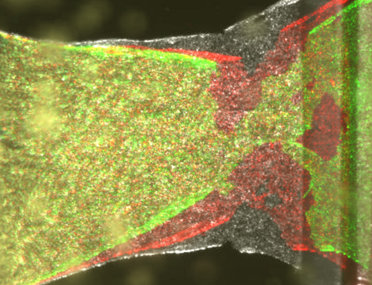
The monolayers can withstand extreme deformations due to the keratin networks that connect cells together and become rigid under stress, preventing the tissues from tearing. However, when the keratin network is interrupted, the tissues lose this protective capacity, highlighting the importance of the network in maintaining the tissue’s integrity. The study also highlighted how epithelial monolayers resist breakage depending on the stretching speed and can expand three to nine times their initial length. The slower they are lengthened, the more they can deform before breaking.
To better understand how epithelial monolayers break, advanced experiments — guided by Julia Duque — were combined with computational models. We discovered that these individual cell layers can stretch up to nine times before they rupture!
Alessandra Bonfanti, Department of Civil and Environmental Engineering, co-author of the study
The monolayers can withstand extreme deformations due to the keratin networks that connect cells together and become rigid under stress, preventing the tissues from tearing. However, when the keratin network is interrupted, the tissues lose this protective capacity, highlighting the importance of the network in maintaining the tissue’s integrity. The study also highlighted how epithelial monolayers resist breakage depending on the stretching speed and can expand three to nine times their initial length. The slower they are lengthened, the more they can deform before breaking.
Tissue rupture is a multiscale process. The model connects events on the molecular scale, such as breakage of the adhesive bond, with the response on the tissue scale when it is stretched. This allowed the researchers to simulate and predict how the tissues behave when the deformation speed changes, revealing how the keratin networks contribute to their strength. Without these models, we would not be able to connect the cellular and molecular processes, or what happens with the tissues.
The work combined knowledge of biology, physics, and engineering, representing an important first step in understanding the complex process of tissue rupture.
Duque, J., Bonfanti, A., Fouchard, J. et al.
Rupture strength of living cell monolayers.
Nat. Mater. 23, 1563–1574 (2024).